The images in this gallery show real cells under the microscope. Do they look like cell diagrams you’ve seen? Probably not!
Most cell diagrams, whether in your textbook or online, are generic. They highlight a set of overlapping features that all cells need to live. But every cell also has unique features to do a specialized job. The examples here explore structures you probably won’t find on most cell diagrams.
Hint: Turn the labels on and off to find the structures on the microscope view.
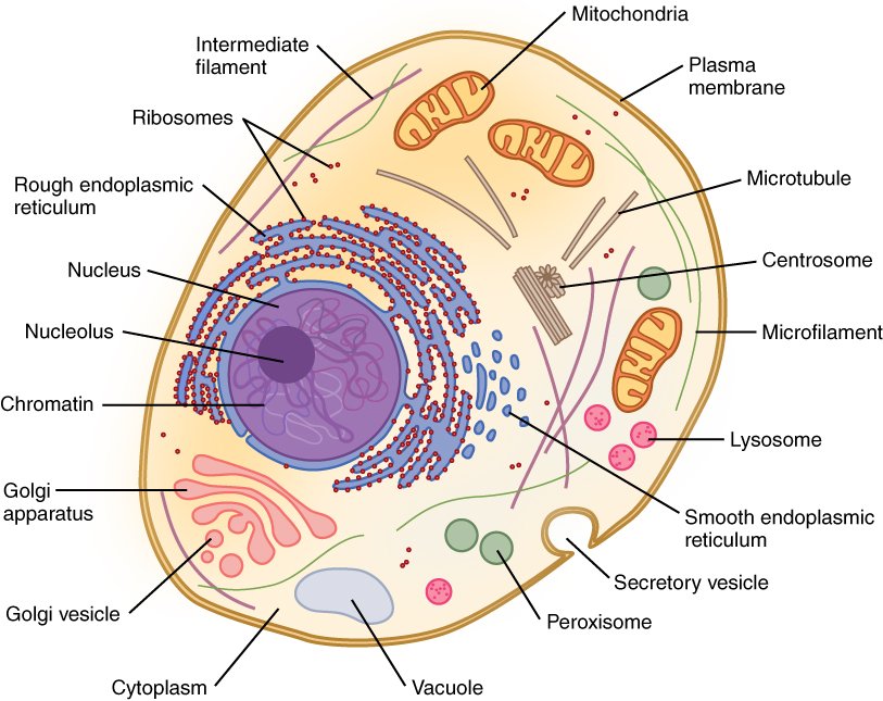
Cell diagrams like this one highlight the parts most cells have in common. Souce: OpenStax via Wikimedia Commons, licensed under CC-BY 4.0.
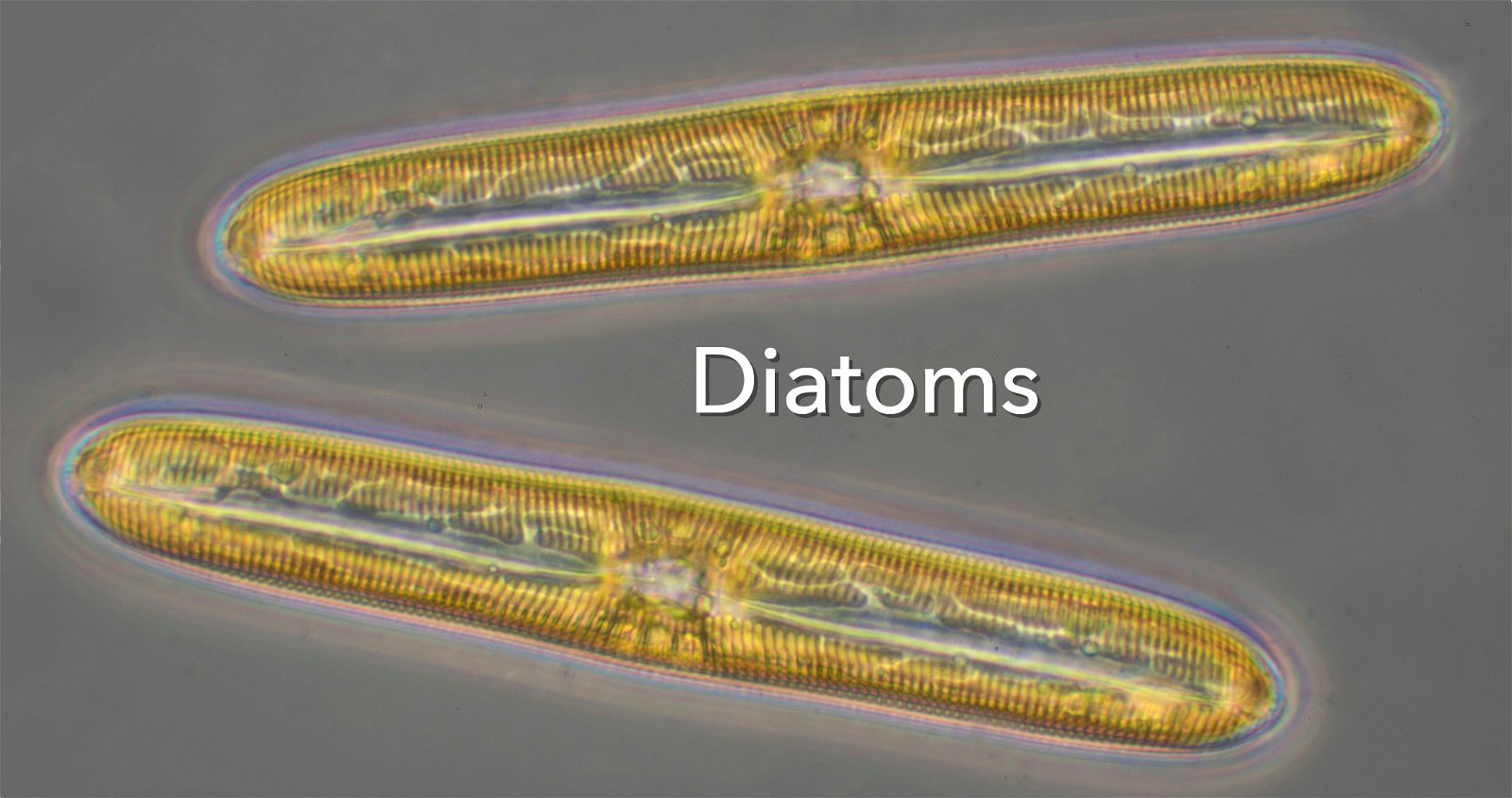
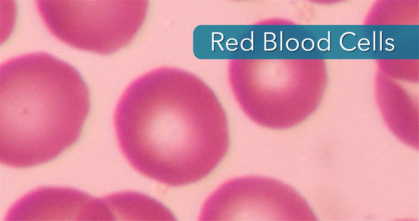
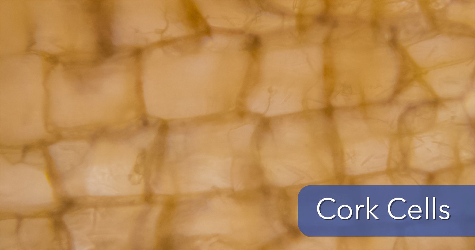
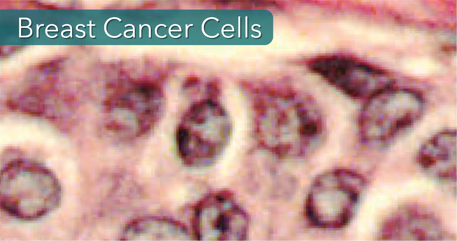
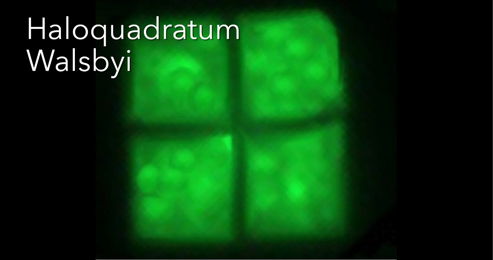
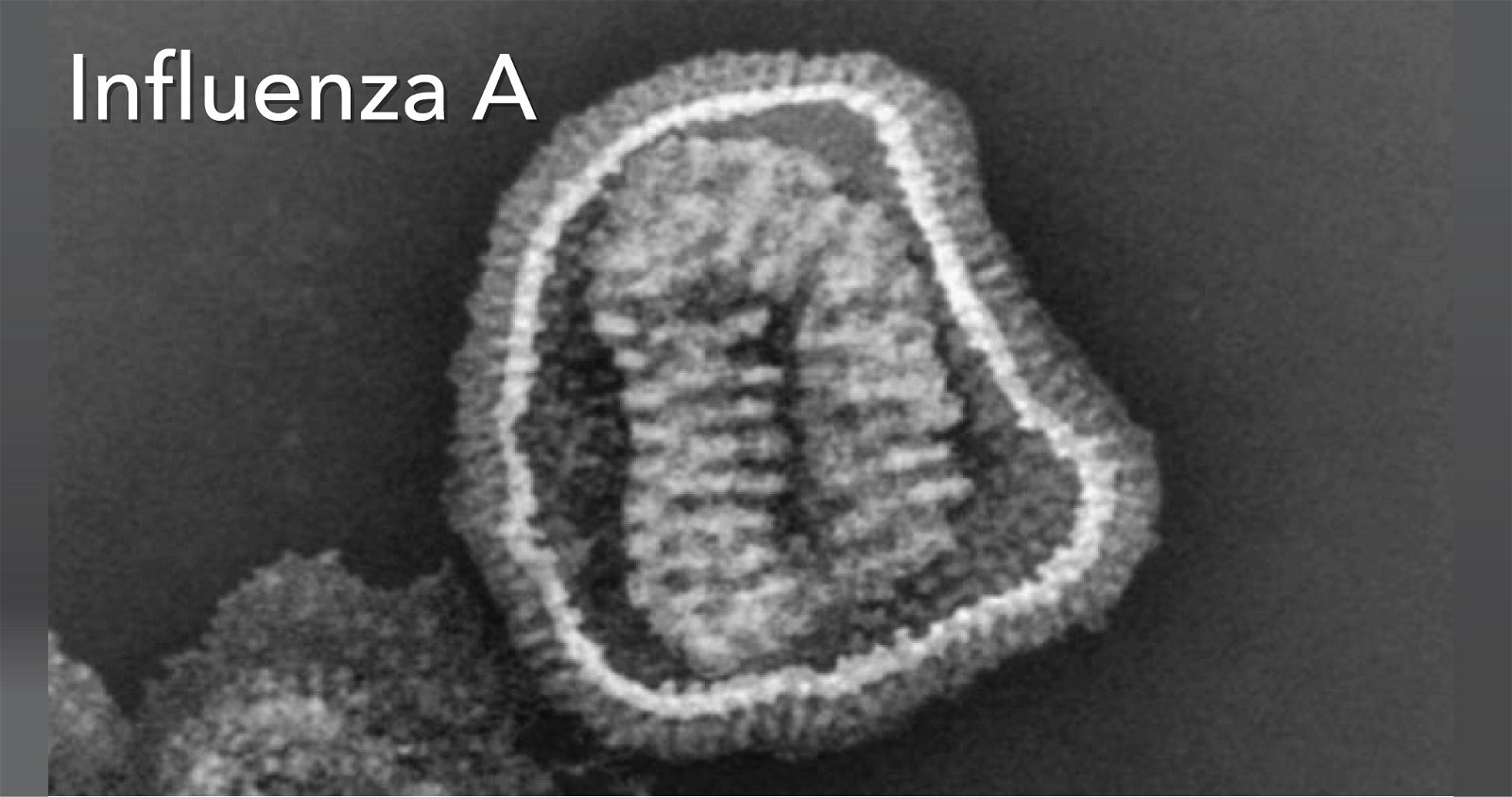
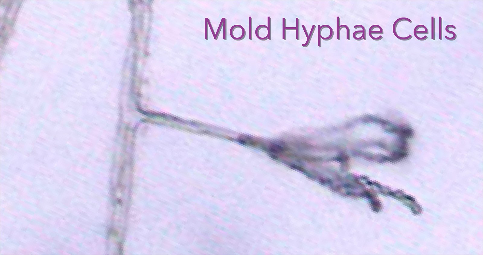
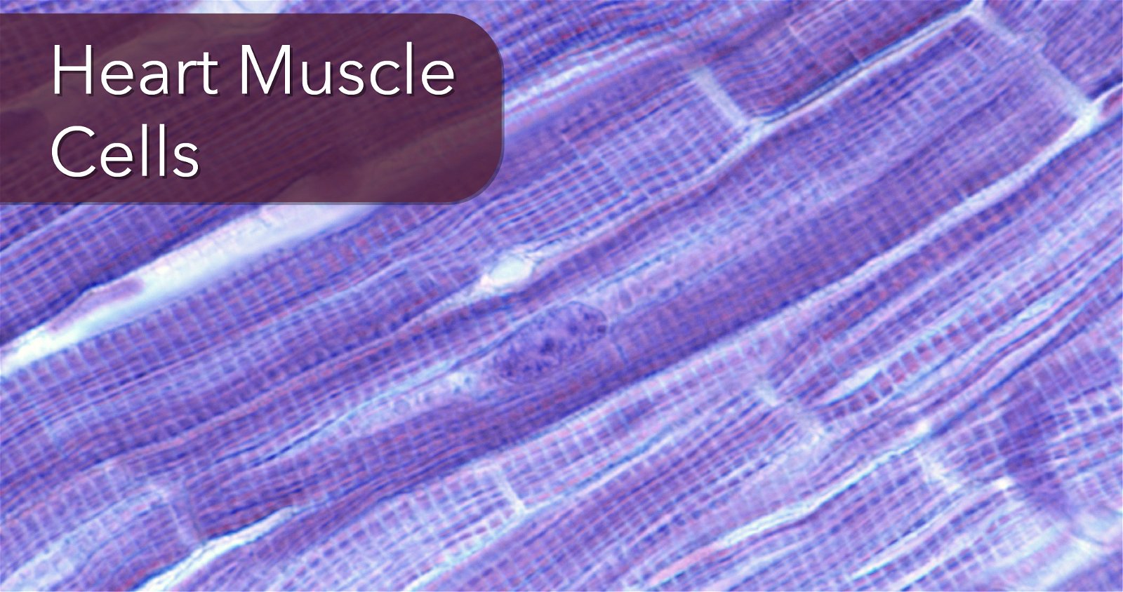
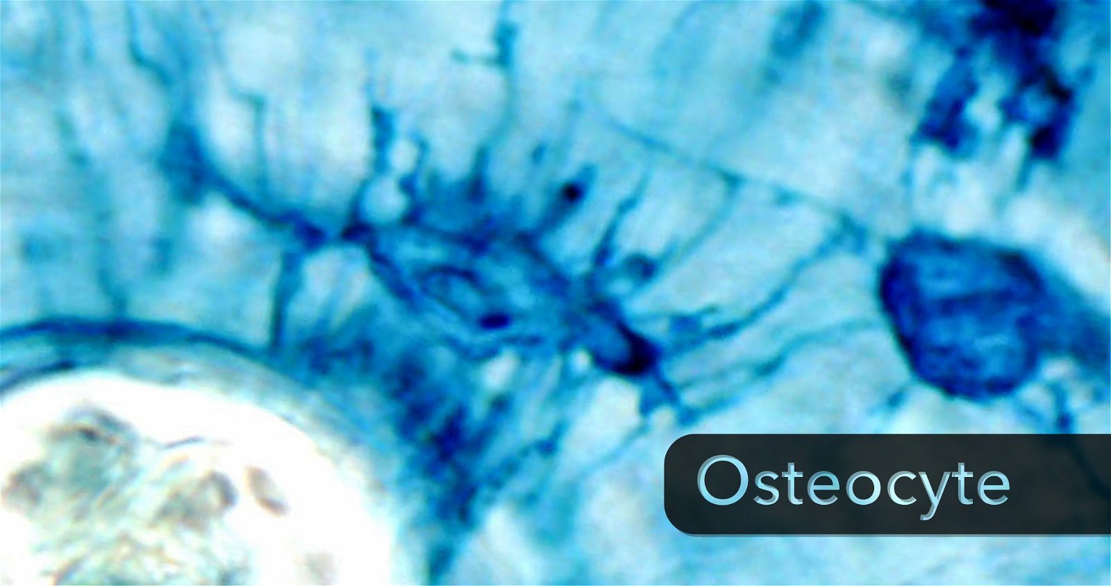
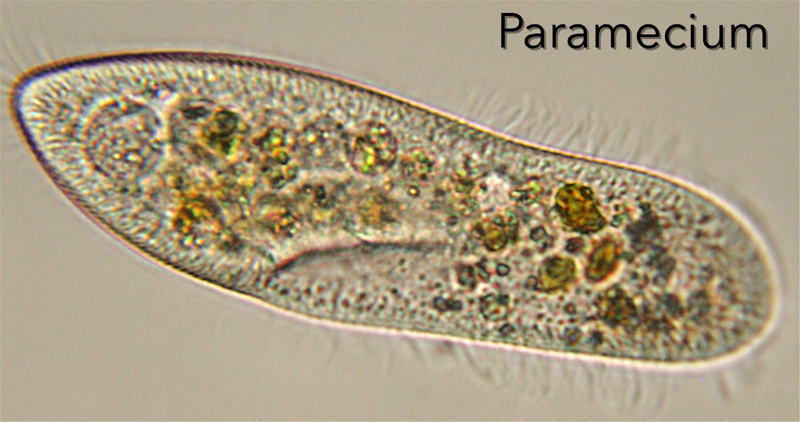
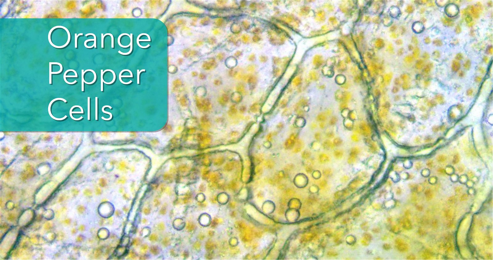
Image Credits
Breast Cancer Cells: National Cancer Institute
Diatom: Robert Pickett
Haloquadratum walsbyi: Mike Dyall-Smith
Heart Muscle Cell: Jose Martin and Jose Enrique Muzquiz
Influenza A: Frederick Murphy, CDC Public Health Image Library
Mold hyphae: Y. Tambe; licensed under CC BY-SA 3.0. Cropped image and added labels.
Osteocyte: Jose Luis Calvo